Flashcards · Biology · Cell Structure
Microscopy and Magnification
-
Write the magnification formula.
Magnification = size of image ÷ size of real object.
-
How do you find the real size from the image size and the magnification?
Real size = image size ÷ magnification.
-
What is resolution?
How clearly two points that are close together can be told apart.
-
Which has higher magnification and resolution: light or electron microscope?
The electron microscope.
-
Name two disadvantages of an electron microscope.
It is large and expensive, and the specimen must be dead.
-
Name an advantage of the light microscope.
It is cheap and easy to use, and living cells can be viewed.
-
A ×10 eyepiece and a ×40 objective lens are used. What is the total magnification?
×400.
-
How many micrometres are in 1 mm?
1000.
-
How many nanometres are in 1 µm?
1000.
-
Why is a stain used on a slide?
It makes the cell structures easier to see.
-
Which objective lens should you start with?
The lowest power one, which gives the widest view.
-
Which wheel do you use to find the image, and which to make it sharp?
The coarse focus wheel finds it, then the fine focus wheel sharpens it.
-
Why is a coverslip lowered at an angle?
To avoid trapping air bubbles.
-
How should a biological drawing be done?
Sharp pencil, clear single lines, no shading, labelled, with the magnification written.
-
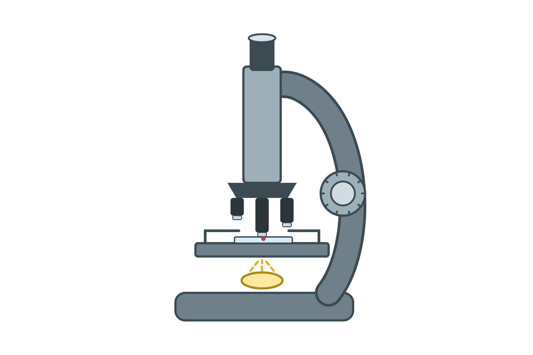 Label every part of this light microscope.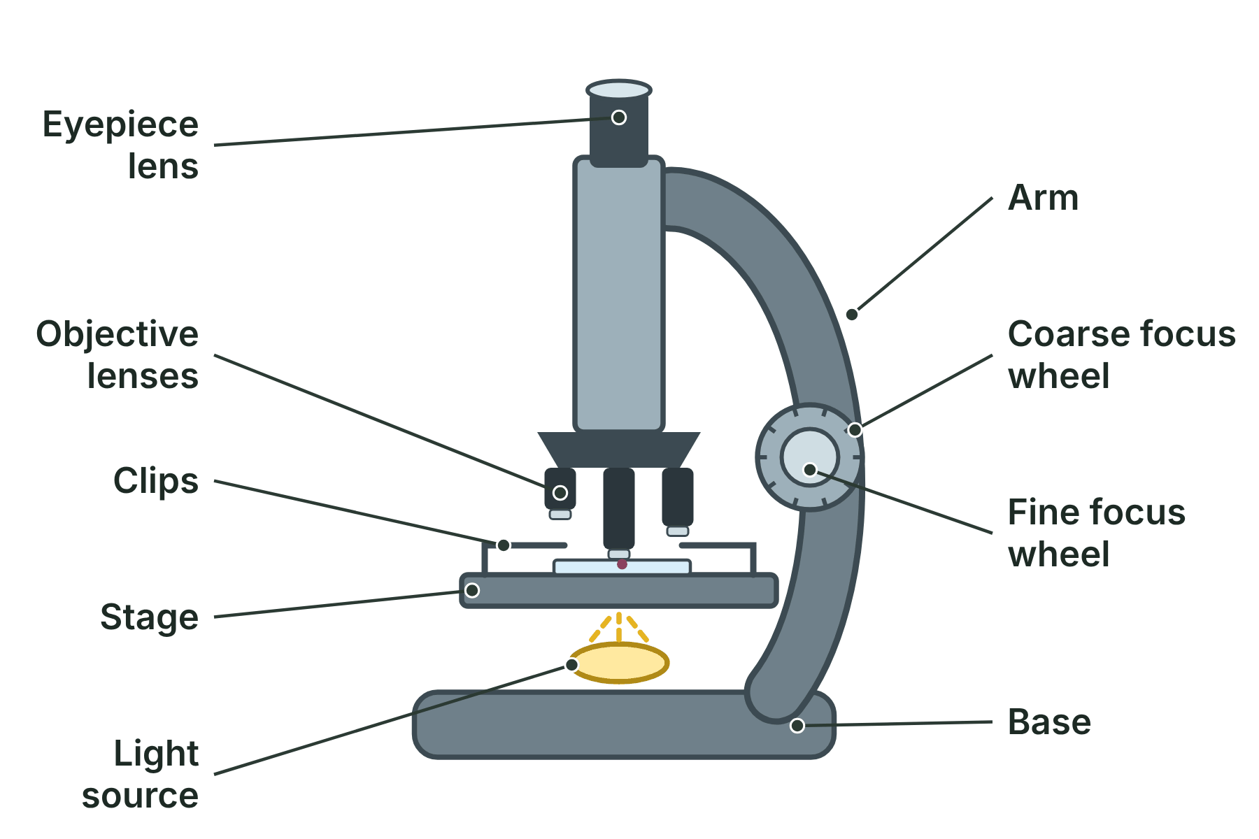
-
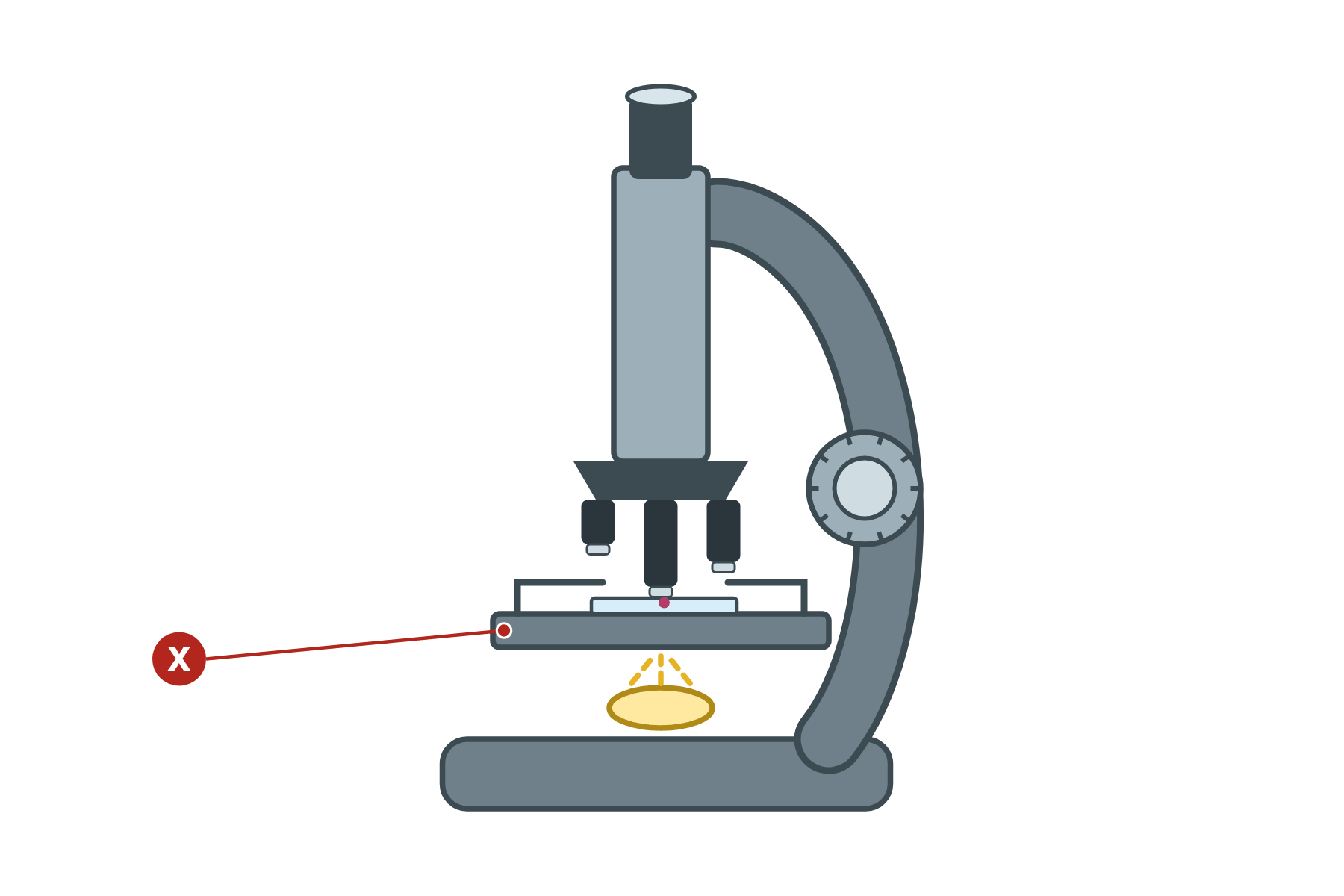 Name part X of the microscope and say what it does.
 The stage. It is the platform that holds the slide, kept in place by the clips.
-
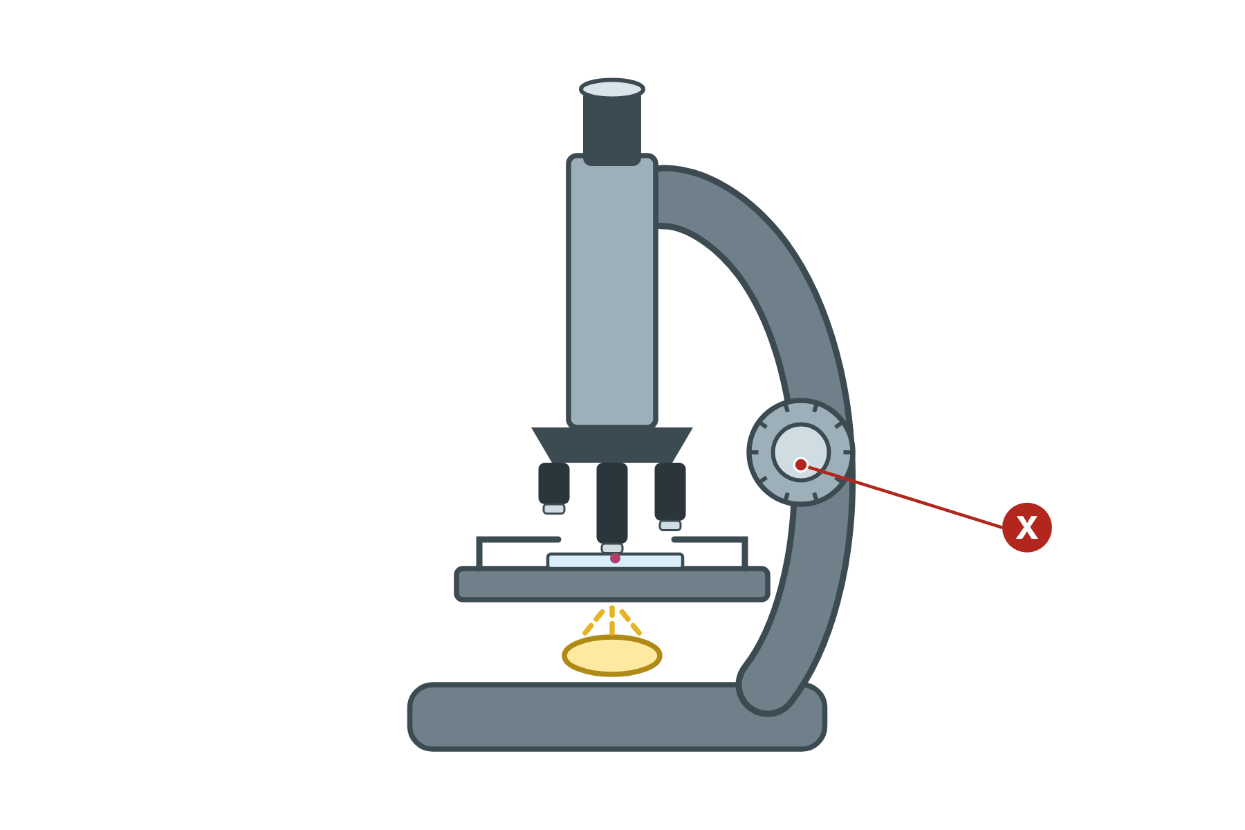 Name part X of the microscope and say what it does.
 The fine focus wheel. It makes small adjustments to give a sharp, clear image, after the coarse wheel has found it.