1.6 Microscopes and magnification
7 exam questions worth 16 marks. Write your answer, open the mark scheme, then give yourself the marks you earned. Your answers are saved on this device.
0 / 16
Which statement about electron microscopes is correct?
Choose one answer.
Correct Not quite: the answer is They have a higher resolution than light microscopes
Mark scheme
C (They have a higher resolution than light microscopes)
Model answer
C: They have a higher resolution than light microscopes. Electron microscopes have a much higher resolution, so they show more detail.
Give two advantages of using an electron microscope instead of a light microscope.
Mark scheme
Any two from:
- higher magnification (1)
- higher resolution / shows more detail (1)
- can show sub-cellular structures such as ribosomes (1)
Model answer
An electron microscope has a higher magnification and a higher resolution, so it shows much more detail, such as ribosomes.
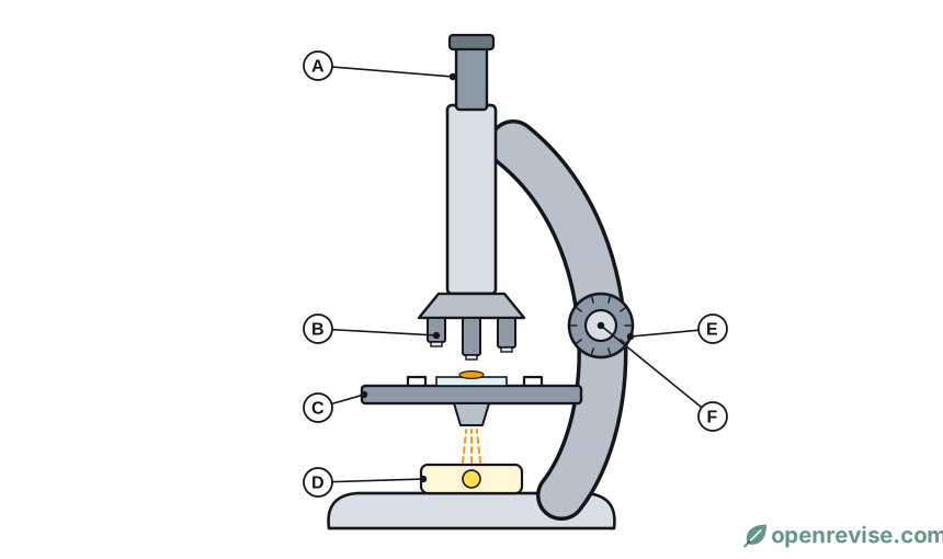
Name the parts labelled A, B and D.
Mark scheme
- A: eyepiece (lens) (1)
- B: objective lens (1)
- D: light source / lamp / mirror (1)
Model answer
A is the eyepiece lens, B is an objective lens and D is the light source.
A student looks at a cell using a ×10 eyepiece lens and a ×40 objective lens.
(a) Calculate the total magnification. (1 mark)
(b) The image of the cell is 3.2 mm long. Calculate the real length of the cell in micrometres (µm). (2 marks)
Mark scheme
(a) 10 × 40 = ×400 (1)
(b)
- 3.2 ÷ 400 = 0.008 (mm) (1)
- = 8 (µm) (1)
- allow 8 µm for 2 marks with no working
Model answer
(a) 10 × 40 = ×400.
(b) real size = image size ÷ magnification = 3.2 ÷ 400 = 0.008 mm. Multiply by 1000 to change to micrometres: 8 µm.
An electron micrograph of a mitochondrion has a magnification of ×20 000. The image of the mitochondrion is 40 mm long.
Calculate the real length of the mitochondrion in micrometres (µm).
Mark scheme
- 40 ÷ 20 000 (1)
- = 0.002 (mm) (1)
- = 2 (µm) (1)
- allow 2 µm for 3 marks with no working
Model answer
real size = 40 ÷ 20 000 = 0.002 mm. Since 1 mm = 1000 µm, 0.002 mm = 2 µm.
Ribosomes can be seen with an electron microscope but not with a light microscope.
Explain why.
Mark scheme
- ribosomes are very small (1)
- the electron microscope has a higher resolution (1)
- so it can show two points that are very close together as separate points (1)
- maximum 2 marks
Model answer
Ribosomes are very small. An electron microscope has a much higher resolution than a light microscope, so it can show details that are too small and too close together for a light microscope to separate.
Methylene blue is added to cheek cells before they are viewed with a light microscope.
Explain why a stain is used.
Mark scheme
- cells are (almost) transparent / colourless (1)
- the stain makes structures such as the nucleus show up / increases contrast (1)
Model answer
Cheek cells are almost transparent. The stain colours structures such as the nucleus so that they show up and can be seen clearly.
Found a mistake?