1.6 Microscopes and magnification
Light and electron microscopes compared, magnification versus resolution, the parts of a light microscope, stains, and how to calculate magnification and real size, including unit conversions.
Seeing cells
Key terms
- Magnification: how many times bigger an image is than the real object.
- Resolution: the ability to see two points that are close together as separate points. A higher resolution shows more detail.
- Light microscope: a microscope that uses light and glass lenses to magnify a specimen.
- Electron microscope: a microscope that uses a beam of electrons instead of light, giving much higher magnification and resolution.
- Eyepiece lens: the lens you look through. Together with the objective lens it makes the total magnification.
- Objective lens: a lens close to the specimen. A microscope has several, of different magnifications.
Cells are too small to see with the naked eye, so biologists use microscopes. Better microscopes have helped us to understand much more about cells.
Light and electron microscopes
| Feature | Light microscope | Electron microscope |
|---|---|---|
| Uses | Light and glass lenses | A beam of electrons |
| Maximum magnification | About ×1500 | About ×2 000 000 |
| Resolution | About 0.2 µm (200 nm) | About 0.2 nm, roughly 1000 times better |
| What you can see | Cells, nuclei, chloroplasts, cell walls | Fine detail inside cells, such as ribosomes and the inner structure of mitochondria |
| Living specimens | Yes | No: the specimen is in a vacuum |
| Cost and size | Cheap, small and easy to use | Very expensive, large, needs trained operators |
| Image | Colour, often with a stain | Black and white (colour can be added later) |
The electron microscope has a much higher resolution than the light microscope, so it can show sub-cellular structures in much finer detail. Using it, biologists discovered structures that are too small to see with a light microscope, such as ribosomes, and understood how mitochondria and chloroplasts work. There are two main types: a transmission electron microscope shows the inside of a thin slice of a specimen, and a scanning electron microscope shows the 3D surface.
Parts of a light microscope
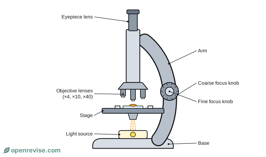
- The eyepiece lens is the lens you look through. It usually magnifies ×10.
- The objective lenses, usually ×4, ×10 and ×40, are on a rotating nosepiece. You choose one to set the magnification.
- The stage holds the slide, and clips keep it in place.
- The light source shines light up through the specimen.
- The coarse focus knob moves the stage up and down to bring the specimen roughly into focus. The fine focus knob makes small changes to get a sharp image.
Total magnification = eyepiece lens magnification × objective lens magnification. For example, ×10 × ×40 = ×400.
Stains
Most cells are almost transparent, so a stain is used to make some parts stand out. Iodine solution stains plant cells brown, and methylene blue stains animal cells such as cheek cells, turning the nucleus blue.
Calculating magnification
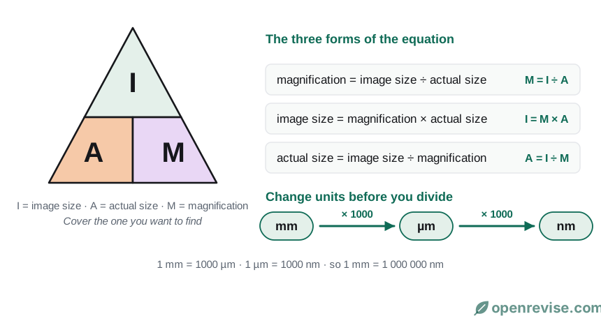
Put the image size and real size in the same units before you divide. Magnification has no units. Remember: mm to µm, multiply by 1000; µm to nm, multiply by 1000.
Worked example 1: finding the real size. A photograph of a cell is 45 mm long. The magnification is ×300.
real size = image size ÷ magnification = 45 ÷ 300 = 0.15 mm = 150 µm
Worked example 2: finding the magnification. An electron micrograph shows a mitochondrion that is 30 mm long. Its real length is 2 µm.
30 mm = 30 000 µm, so magnification = 30 000 ÷ 2 = ×15 000
Test yourself on this lesson
7 exam questions · 16 marks · with mark schemes and model answers
Found a mistake?