5.7 Structure of the eye
The parts of the eye and their jobs: cornea, iris, pupil, lens, ciliary muscles, suspensory ligaments, retina (rods and cones), sclera and optic nerve, plus the iris reflex.
The eye as a sense organ
Key terms
- Retina: the layer at the back of the eye containing light-sensitive receptor cells (rods and cones).
- Cornea: the transparent front of the eye that refracts (bends) light as it enters.
- Lens: a transparent, flexible structure that focuses light onto the retina.
- Iris: the coloured ring of muscle that controls the size of the pupil, and so how much light enters.
- Rods: receptor cells that work in dim light and see in black and white.
- Cones: receptor cells that need bright light and detect colour.
The eye is a sense organ. Its receptors, in the retina, are sensitive to light intensity and colour. The rest of the eye is built to get a sharp image onto those receptors, and to protect them.
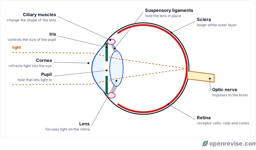
Structures and their functions
| Structure | What it is like | Function |
|---|---|---|
| Cornea | Transparent, curved, no blood vessels | Refracts light into the eye (does most of the bending) |
| Iris | Coloured ring of circular and radial muscles | Controls the size of the pupil and so the amount of light reaching the retina |
| Pupil | A hole in the middle of the iris | Lets light into the eye |
| Lens | Transparent, flexible, held in place | Changes shape to focus light onto the retina |
| Ciliary muscles | A ring of muscle around the lens (part of the ciliary body) | Contract or relax to change the shape of the lens |
| Suspensory ligaments | Strong fibres linking the ciliary muscles to the lens | Hold the lens in place and pull on it |
| Retina | Layer of light receptor cells at the back | Detects light; sends impulses along the optic nerve |
| Optic nerve | Bundle of sensory neurones | Carries impulses from the retina to the brain |
| Sclera | Tough, white outer layer | Protects the eye; muscles that move the eye attach to it |
How we see
- Light enters the eye through the cornea, which refracts it.
- It passes through the pupil, and the lens refracts it further to focus it on the retina.
- The image on the retina is upside down and very small.
- Receptor cells in the retina are stimulated and send impulses along the optic nerve (sensory neurones) to the brain.
- The brain interprets the impulses, so we see the image the right way up.
Nothing travels out of the eye when we look at something: light comes in from objects, and the brain builds the picture.
Rods and cones
The retina contains two kinds of light receptor cell:
- Rods are very sensitive to light, so they work in dim light, but they only give a black-and-white image. They are spread around the edge of the retina, which is why you see faint stars better out of the corner of your eye.
- Cones need bright light and detect colour. There are three types, sensitive to red, green and blue light. They are packed most closely in the middle of the retina (the fovea), where we see in most detail.
If one type of cone does not work properly, a person cannot tell some colours apart: this is colour blindness.
The iris reflex: adjusting to light
The iris contains two sets of muscles: circular muscles in a ring around the pupil, and radial muscles running outwards like the spokes of a wheel. They work against each other.
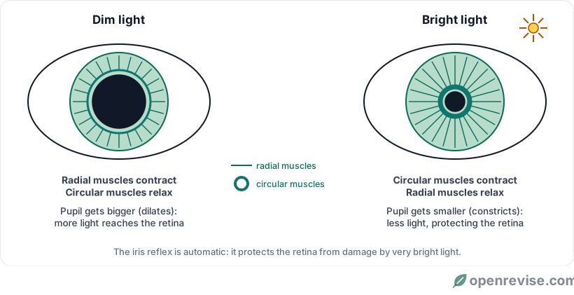
| Part of the eye | Dim light | Bright light |
|---|---|---|
| Circular muscles | Relax | Contract |
| Radial muscles | Contract | Relax |
| Pupil | Gets bigger (dilates) | Gets smaller (constricts) |
| Result | More light enters, so we can see | Less light enters, protecting the retina from damage |
This is a reflex: light receptors in the retina detect the change, and impulses pass through a reflex arc (via the brain, but not the conscious part) to the iris muscles, the effectors.
Test yourself on this lesson
6 exam questions · 15 marks · with mark schemes and model answers
Found a mistake?