5.6 Investigating and treating the brain
How scientists map brain function (patients with brain damage, electrical stimulation, MRI, CT and PET scans) and why brain and spinal injuries are so hard to treat.
Mapping the brain
Key terms
- MRI scan: magnetic resonance imaging. Strong magnets and radio waves make detailed images of the brain without radiation; functional MRI (fMRI) shows which areas are active.
- CT scan: computerised tomography. X-rays taken from many angles are combined to give images of slices through the brain.
- PET scan: positron emission tomography. A radioactive tracer is injected; the scan shows which areas take it up most, so which are most active.
Neuroscientists have linked regions of the brain to particular functions in several ways.
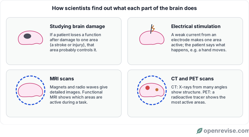
1. Studying patients with brain damage. If one area of the brain is damaged, by a stroke, a tumour or an injury, and the patient loses one ability (such as speech, or movement on one side), that area is probably involved in that ability. A famous case is Phineas Gage, a railway worker whose front brain was pierced by an iron rod in 1848: he survived, but his personality changed. Cases like his linked the front of the cerebrum to personality and decision making.
2. Electrically stimulating the brain. During brain surgery, the patient may be awake (the brain has no pain receptors). Surgeons pass a weak electric current through a small electrode into one area. The patient might twitch a finger, see a flash or recall a memory. This maps which area does what, and helps surgeons avoid damaging vital areas.
3. MRI scanning. Strong magnetic fields and radio waves produce very detailed images of soft tissue. Functional MRI (fMRI) shows which areas have increased blood flow while a person does a task, such as reading or moving a hand, so it shows which areas are active.
4. CT and PET scanning.
| Scan | How it works | What it shows | Drawbacks |
|---|---|---|---|
| CT | Many X-ray images from different angles, combined by a computer | Structure: tumours, bleeding, damage | Uses ionising X-rays; shows structure, not activity |
| PET | A radioactive tracer (usually glucose) is injected; active areas use more glucose | Activity: which areas are working; tumours and diseases such as Alzheimer's | Uses a radioactive substance; expensive; less detailed images |
| MRI | Strong magnets and radio waves | Very detailed structure; fMRI shows activity | Expensive; noisy; cannot be used with some metal implants |
Why investigating the brain is difficult
- The brain is very complex: billions of neurones with trillions of connections, and most functions (memory, language) involve several areas at once.
- It is delicate and hard to reach inside the skull, so tissue cannot simply be removed and studied in a living person.
- Case studies of brain damage are rare and each one is different, so results are hard to repeat and to interpret. The damage may also affect several areas.
- There are ethical issues: you cannot damage a healthy brain to test an idea, and patients with brain damage may not be able to give informed consent.
Why treating brain and nerve damage is difficult
- Neurones in the CNS have a very limited ability to repair or regrow. Damage is often permanent.
- Spinal injuries: if the spinal cord is cut, impulses cannot pass the damage, so the person may lose movement and feeling below the injury (paralysis). The neurones cannot regrow across the gap.
- Brain tumours: surgery to remove a tumour may damage healthy tissue around it, which cannot be replaced, so the patient may lose abilities. Some tumours are too deep to reach safely.
- Access: many areas are deep inside the brain or protected by bone, and many drugs cannot pass from the blood into brain tissue.
- Side effects: treatments such as radiotherapy and chemotherapy can damage healthy neurones too.
Doctors must weigh the benefits (removing a tumour, stopping seizures) against the risks (loss of speech, movement or memory, infection). Patients need clear information to make that choice.
Test yourself on this lesson
6 exam questions · 19 marks · with mark schemes and model answers
Found a mistake?