4.8 Uses and hazards of nuclear radiation
How the type of radiation and the half-life of an isotope decide its use: medical tracers, radiotherapy, sterilising, smoke alarms and PET scans, and the hazards that must be managed. For Separate Science.
Using nuclear radiation
Key terms
- Tracer: a radioactive substance that is put into the body or a system so that its movement can be followed with a detector.
- Radiotherapy: the use of ionising radiation to kill cancer cells.
- Sterilise: to kill all bacteria and other microorganisms, for example on surgical instruments.
- Positron emission tomography (PET): a scan that uses a radioactive isotope that emits positrons to produce an image of the inside of the body. (Edexcel)
Nuclear radiation is dangerous, but it is also very useful. To decide which isotope to use for a particular job, you need to think about two things:
- the type of radiation it emits (alpha, beta or gamma) and how penetrating it is;
- its half-life: it must last long enough to do its job, but not stay radioactive for longer than necessary.
Medical tracers
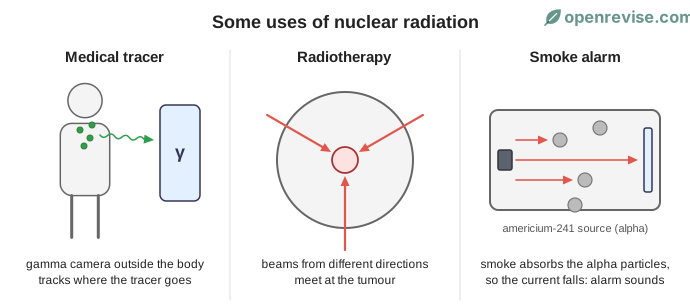
A tracer is a small amount of radioactive material that is injected into the body or swallowed. A gamma camera outside the body detects the gamma rays that are emitted and shows where the tracer has gone. This can be used to check how well organs such as the kidneys, thyroid or heart are working, or to find blockages.
A good medical tracer must:
- emit gamma rays (or another penetrating radiation), so the radiation can leave the body and be detected, and not be strongly ionising inside the body, as alpha would be;
- have a short half-life, a few hours, so it lasts long enough for the scan but decays quickly so the patient's dose is small;
- be non-toxic, and be removed from the body quickly.
An example is technetium-99m, which emits gamma rays and has a half-life of 6 hours.
Radiotherapy
Radiotherapy uses radiation to kill cancer cells.
- External radiotherapy: a beam of gamma rays (for example from cobalt-60) is aimed at the tumour from outside the body. The source is moved around the patient so the beams cross at the tumour. The tumour gets a high dose, but the healthy tissue is only exposed for a short time from any one direction.
- Internal radiotherapy: a small radioactive source, often a beta emitter, is placed inside or next to the tumour. Because beta radiation travels only a short distance in the body, it mainly damages the tumour. A short half-life means that the dose is delivered quickly. Iodine-131 is used for thyroid cancer.
Radiotherapy also damages healthy cells, which is why patients can feel unwell, but the benefit of curing the cancer is greater than the risk.
Sterilising equipment and food
Gamma rays are used to kill bacteria on surgical instruments and on some food. The items are sealed in their packaging first, so they stay sterile afterwards. Gamma rays are used because they are so penetrating that they can pass through the packaging. The items are irradiated, but they do not become radioactive. The source has a long half-life (cobalt-60 has 5.3 years), so it does not need to be replaced often.
Smoke alarms
A smoke alarm contains a small source of americium-241, an alpha emitter. The alpha particles ionise the air in a small gap between two plates, so a small current flows. When smoke enters the gap, it absorbs the alpha particles, the current falls, and the alarm sounds.
Alpha is used because it is absorbed by the smoke particles but is stopped by air or by the alarm's case, so it is safe for people in the room. It has a very long half-life (432 years), so the alarm works for many years without a change of source.
PET scans (Edexcel)
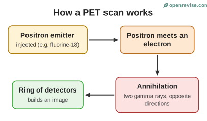
A PET scan uses an isotope that emits positrons, such as fluorine-18 (half-life about 110 minutes), attached to a glucose-like molecule. Active cells take up more of it, for example brain cells or tumours. Each positron meets an electron and both are destroyed, giving two gamma rays that travel in opposite directions. A ring of detectors picks them up to build a 3D image.
Choosing the right isotope
| Use | Radiation needed | Half-life needed |
|---|---|---|
| Medical tracer | Gamma (penetrates the body) | A few hours |
| External radiotherapy | Gamma (penetrates to the tumour) | Years (so the source lasts) |
| Internal radiotherapy | Beta (short range) | Hours to days |
| Sterilising | Gamma (penetrates packaging) | Years |
| Smoke alarm | Alpha (absorbed by smoke, safe in a case) | Hundreds of years |
Hazards
All of these uses involve a risk: ionising radiation can damage healthy cells and cause cancer. The risk is made as small as possible by limiting doses, shielding, and using the shortest half-life that will do the job. The benefits of the use must be greater than the risks.
Test yourself on this lesson
9 exam questions · 27 marks · with mark schemes and model answers
Found a mistake?